Biology Laboratories
The Microbiology Prep Room is located in room 531A Life Science Building at Bowling Green State University. The facility supports both undergraduate classes and research laboratories in the Biological Sciences Department.
The Prep Room maintains records as required by the Ohio EPA for the storage, treatment, and disposal of Infectious waste. All infectious waste produced by the biology, chemistry and other departments on campus are treated here.
Link to infectious waste disposal procedures.
For more information, contact Sheila Kratzer, Microbiology Technician, at skratze@bgsu.edu.
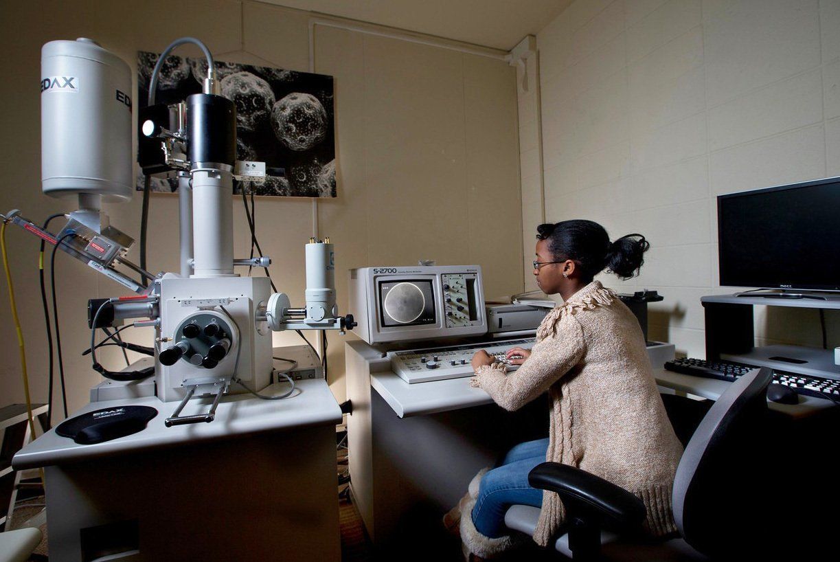
The Scanning Electron Microscope (SEM) is used in many fields of science. A biologist might use it to study the tiniest structures of an insect, the geologist might use it to learn what chemicals are present in a rock, and the automobile engineer might use it to find tiny defects in a car part. SEM images are created using electrons instead of the photons of light we use to see the world around us. Electrons have a shorter wavelength than photons allowing much greater magnifications and resolution than with conventional light microscopy.

A Transmission Electron Microscope (TEM) is used if even higher magnifications are needed. A biologist might use it to study the membrane of an organelle. The chemist might use it to identify a crystalline substance. A crystalline structure can be determined by studying the patterns of electron diffraction made when the electron beam goes through an ultra-thin crystal.

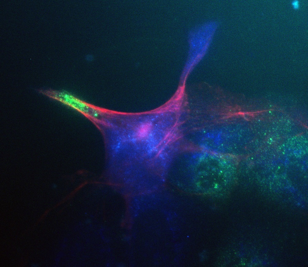
Photons help us view the microscopic world, as for example, in the familiar light microscope and the Confocal Scanning Light Microscope. The confocal microscope captures images at different depths in a thick material, prepared as living or non-living (fixed) tissue.

Welcome to our archive of 185+ digitized images. Note: Any image may be freely used for educational and noncommercial purposes, however, we do reserve all commercial rights.
Sections
Updated: 12/06/2021 06:49PM
