Microscopy and Microanalysis
Microscopy and Imaging Facility
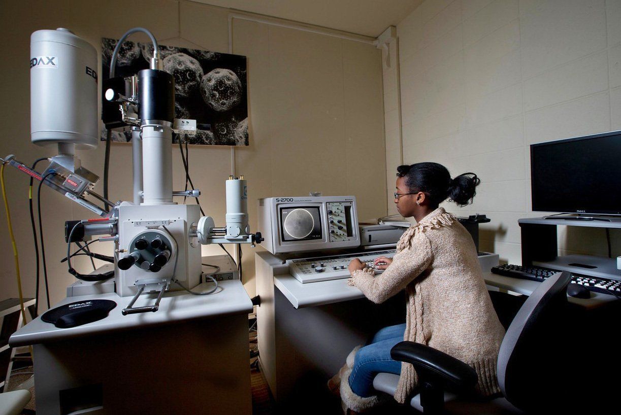
Fluorescence and electron microscopes are available for use by student and faculty researchers.
See the Director Marilyn Cayer for more information.
The Facility is located on the 5th floor of the Life Sciences Building.
The Scanning Electron Microscope (SEM) is used in many fields of science. A biologist might use it to study the tiniest structures of an insect, the geologist might use it to learn what chemicals are present in a rock, and the automobile engineer might use it to find tiny defects in a car part. SEM images are created using electrons instead of the photons of light we use to see the world around us. Electrons have a shorter wavelength than photons allowing much greater magnifications and resolution than with conventional light microscopy.

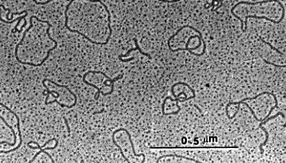
A Transmission Electron Microscope (TEM) is used if even higher magnifications are needed. A biologist might use it to study the membrane of an organelle. The chemist might use it to identify a crystalline substance. A crystalline structure can be determined by studying the patterns of electron diffraction made when the electron beam goes through an ultra-thin crystal.

Light microscopy
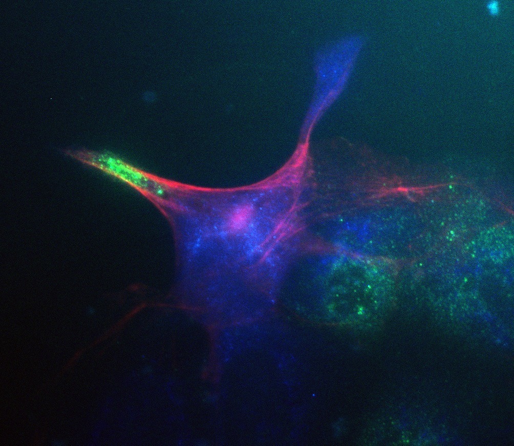
Zeiss and Leica epifluorescence microscopes are available including a spinning-disk confocal microscope suitable for studies of live cells and organisms. Confocal microscopes capture images at different depths in a thick material, prepared as living or non-living (fixed) tissue. Light from a single focal plane is captured to increase image sharpness.
LED light engines and Metamorph imaging software are used along with semiautomated control of the confocal microscope.
Dissecting microscopes equipped with digital cameras are also available.

Updated: 02/25/2022 03:30PM
